Using AI to improve the lives of cancer patients
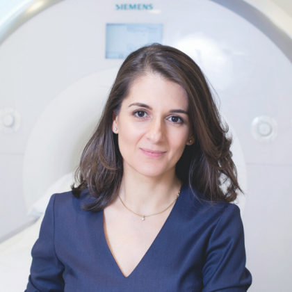
Q&A with consultant radiologist Dr Christina Messiou, who is leading a study to harness Artificial Intelligence (AI).

What is the aim of this study?
In collaboration with Imperial College London, the MALIMAR (Machine Learning in Myeloma Response) study is using machine learning, a type of AI in which computers are taught how to do things independently, to read whole‑body MRI scans in myeloma patients to find evidence of cancer.
What is a whole-body MRI scan?
Unlike CT scans, whole‑body MRI scans can detect cancer in the bone marrow before it has caused destruction to the outer bone, meaning a diagnosis can be made much earlier.
This is particularly important for patients with myeloma, a blood cancer that originates in the bone marrow. As the disease progresses, it can result in irreparable bone damage, leading to debilitating complications for patients. By diagnosing myeloma as early as possible, it can avoid bone damage and vastly improve patients’ quality of life.
Whole body MRI has been recommended by NICE as first line imaging for all patients with myeloma in the UK since 2016. Although, to date not as many centres have implemented it as we would like.

What difference could this mean to patients in the future?
By developing AI to assist radiologists, we will detect and quantify the amount of disease faster and more accurately, allowing patients to start treatment sooner and avoid long-term side effects.
We are also taking steps to make whole-body MRI scans more widely available throughout the UK so that all patients with suspected myeloma, or who are undergoing treatment for myeloma, can benefit regardless of where they live. These steps include the development of AI, but also helping to train radiologists in how to perform and report whole body MRI scans, with 250 radiologists having attended our courses so far.
Dr Christina Messiou is part-funded by The Royal Marsden Cancer Charity.

